CT Scans and PET Scans

Katherine Garner • November 8, 2022

CT Scans and PET Scans


written by
Katherine Garner


PET and CT scans are both important imaging tools, but you might not know the differences between them. This article will explain what PET and CT are, how they produce images, and in which situations each scan may be used.


What are PET and CT? 


PET and CT stand for Positron Emission Tomography and Computed Tomography, respectively. Tomography - with roots in the Greek tomos, meaning ‘section’ or ‘slice’ - is the process of imaging the inside of the body in a series of cross-sectional slices. These slices are digitally stacked to recreate a three-dimensional image of the patient, organ, or tissue. PET and CT both create these slices, but they use different technologies to do so, providing different medical information as a result. 


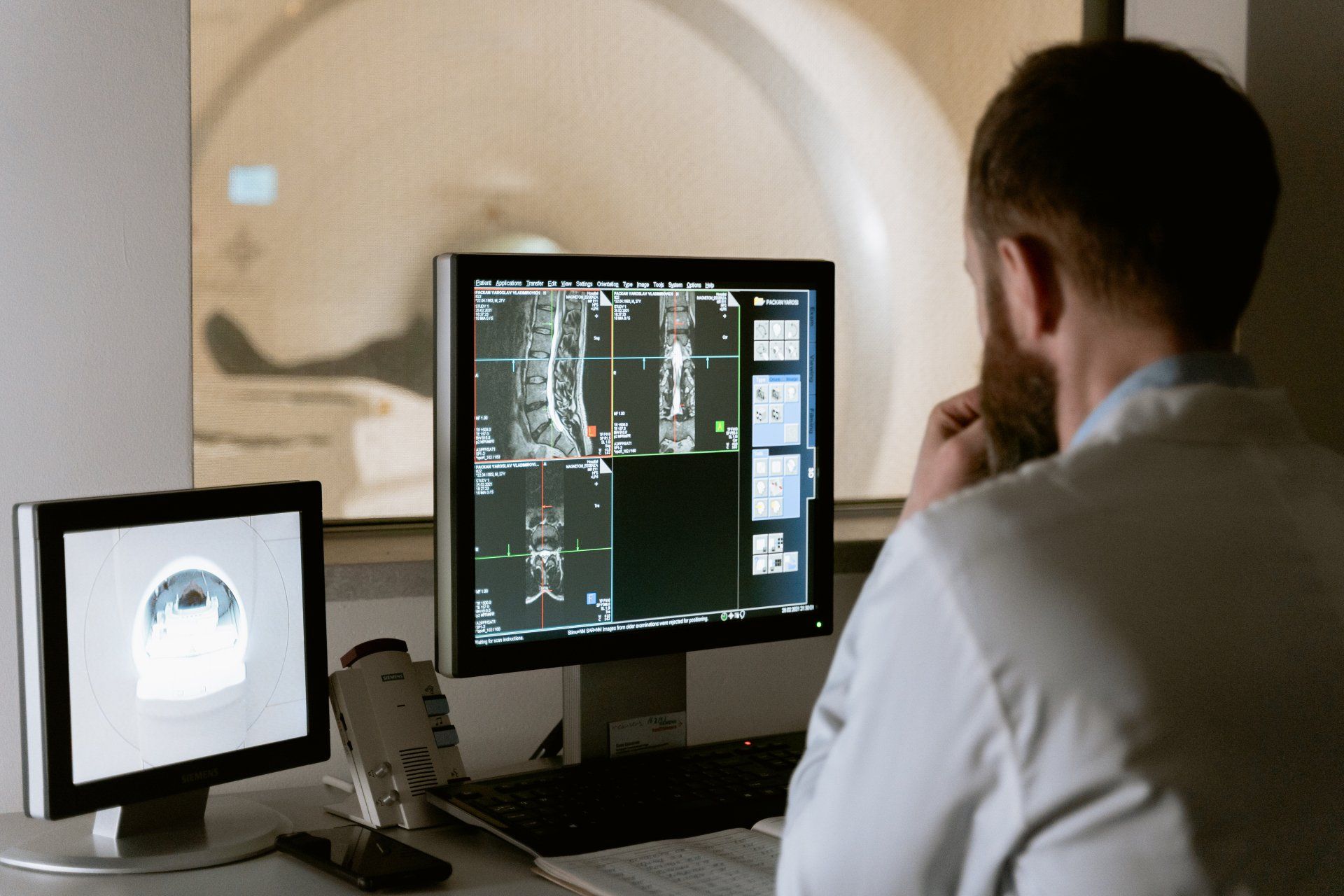
CT uses x-ray radiation to image the inside of the body. In a CT scanner, the patient is flanked by an x-ray beam on one side, and a detector on the other. Unlike a conventional x-ray, CT is three-dimensional: the x-ray beam and detector rotate around the patient to capture the same section from different angles. The amount of x-ray radiation which reaches the detector depends upon the density of the body part it is passing through. By analysing the amount of radiation detected at different positions in the rotation around the patient, a computer programme can reconstruct an internal image of the body. 


Like CT, PET is a three-dimensional scan. Instead of using energy from x-rays, a PET scanner detects tiny spikes of radiation produced by positrons. Positrons are small particles which are given off when an injected radioactive drug, called a radiotracer, decays over time. When these positrons collide with body tissue, they produce spikes of radiation. The scanner detects these spikes and converts them into an image showing where the radiotracer has accumulated. 


PET radiotracers are often radioactive versions of substances used by the body, such as sugar, amino acids, or ammonia: this enables them to be taken up or used by different tissues. Different radiotracers are designed to accumulate in different tissues and cause them to become visible to the scanner.


The most commonly used radiotracer in oncology is a sugar called [18F]-fluorodeoxyglucose, or [18F]-FDG. Cells can take up natural sugars like glucose and break them down for energy, so they can take up [18F]-FDG too. However, this sugar is chemically modified so that it cannot be broken down and accumulates in the cell. Very energy-intensive cells which use lots of glucose, like cancer cells, will also accumulate more [18F]-FDG than other cells. This makes tumours appear as activity ‘hotspots’ on the PET scan.


What information can PET and CT tell us?


By using different radiotracers, PET can be tailored to show different information about the patient. We have talked about the tumor radiotracer [18F]-FDG, but there are many others:


  • Choline is a nutrient which participates in the production of new cell membranes. Areas where many new cells are being produced, such as tumors, will therefore take up more choline than normal cells. Radiolabeled choline can be more accurate than [18F]-FDG when imaging tumors in energy-intensive organs like the brain.
  • [82Rb]-Rubidium chloride is absorbed from the blood very effectively by cells in the heart muscle (myocardium). It can therefore be used to identify areas of the heart which are not receiving enough blood; this is called a myocardial perfusion scan.
  • [18F]-Sodium fluoride is a salt which is used to image bone diseases. It adheres to the outer layer of the bones, acting similarly to the protective coating that fluoride toothpaste forms on teeth.


CT is used to obtain realistic images of the internal organs. Unlike a normal two-dimensional x-ray, CT offers precise spatial information which may be required in certain cases. It offers a non-invasive way to identify sites of damage after a major traumatic injury, for example, and shows soft tissues better than a normal x-ray. CT can be combined with further contrast-enhancing agents, for example intravenous dyes which make blood vessels more visible in the scan; this can be used to locate abnormalities like aneurysms or blood clots.


In some cases, both PET and CT may contribute different useful information, and in this case a combined PET-CT may be used.


Who can have a CT or PET scan?


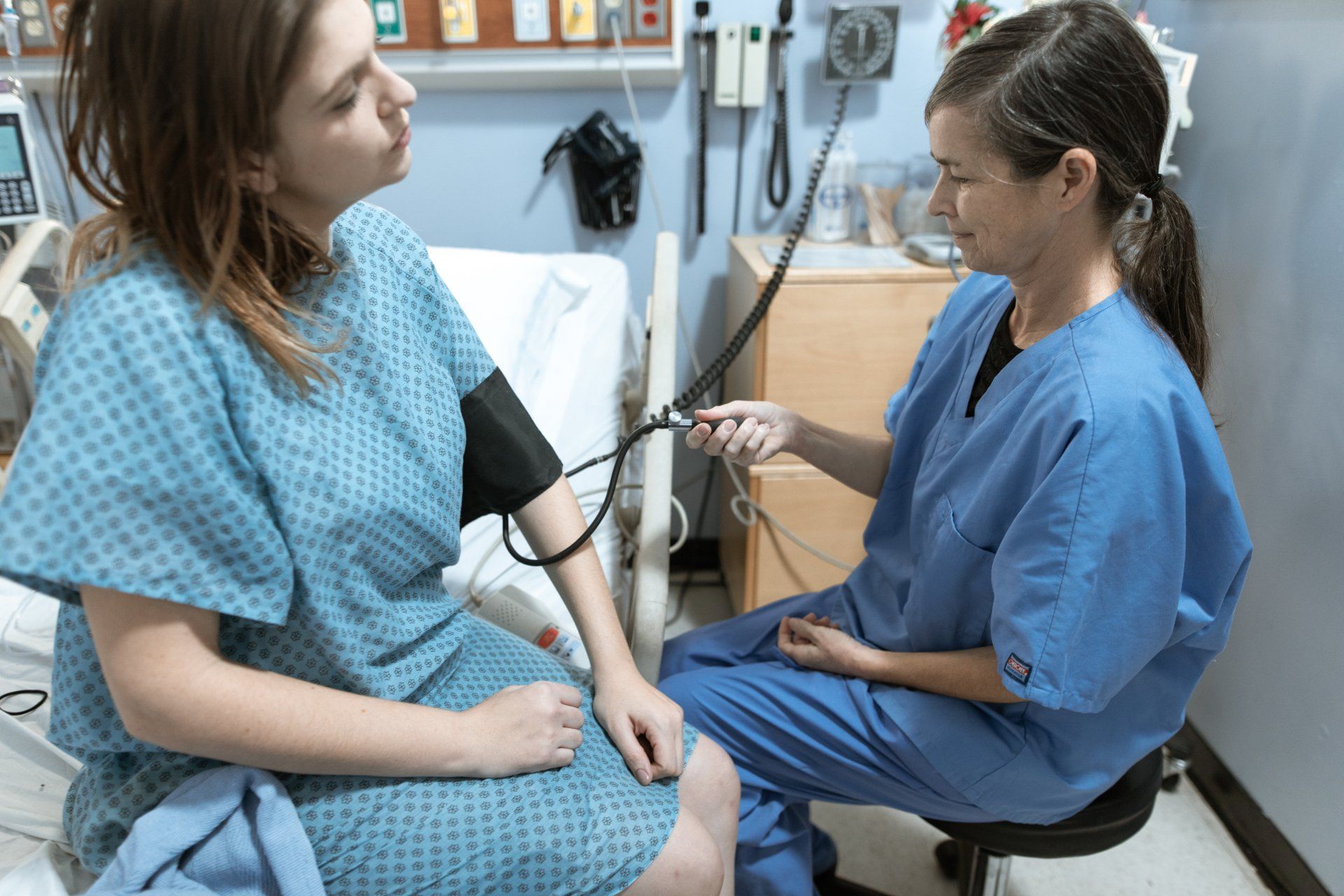
Unlike Magnetic Resonance Imaging (MRI), both PET and CT are safe for use in patients with pacemakers or metal implants. However, radiation dose is an important safety consideration.


Radiotracers are considered safe because they lose their radioactivity rather quickly; this is why they are injected into the patient no more than an hour before a PET scan. The radiotracer is degraded and flushed out of the body within a few hours.

Researchers have estimated that a whole-body combined PET/CT scan exposes the patient to a dose of around 12 millisieverts, which is three times the yearly radiation exposure of the average person (a useful visualisation of these doses can be found here). Hence, the use of these procedures is not a decision to be taken lightly. The safety of PET and CT may be improved further using technologies which reduce the required radiation dose, such as machines with image enhancement algorithms.


Usually, PET and CT are avoided in pregnant women and babies as a precautionary measure. In some cases, however, they might be necessary, for example to locate a tumor for treatment which could negatively affect the pregnancy if left untreated. Locating the tumor by PET or CT exposes the fetus to radiation, but the risk to mother and child of leaving the tumor untreated might be far greater.


Summary


  • CT and PET are both types of tomography – an imaging method which constructs 3D images of the body from a series of "slices".
  • CT provides anatomical images of the body, whereas PET provides information about the activity and function of different tissues.
  • Each scan uses a form of radiation. CT uses external X-rays which are passed through the patient, whereas PET uses radiotracers which can be taken up by different tissues in the body.


Sources

CT Scan - National Health Service

How Does it Work? Positron Emission Tomography - British Medical Journal

PET Scan - National Health Service

Brain tumour imaging with carbon-11 choline: comparison with FDG PET and gadolinium-enhanced MR imaging - European Journal of Nuclear Medicine

Myocardial Perfusion Scan - StatPearls

Clinical PET Imaging in Prostate Cancer - RSNA Radiographics

Radiation Dose from Whole-Body F-18 Fluorodeoxyglucose Positron Emission Tomography/Computed Tomography: Nationwide Survey in Korea - Journal of Korean Medical Science

Radiation Dose Chart - xkcd

The use of PET/CT in pregnancy: A case report of malignant parathyroid carcinoma and a review of the literature - Obstetric Medicine


Katherine is currently completing a Masters by Research in Cancer Biology at Imperial College London. She is enthusiastic about science communication and is a contributor to both of the College’s science magazines, I, Science and Broadsheet, where further examples of her writing can be found. She can be contacted on LinkedIn.


Thank you for reading Patient Education Essentials, the Write Shift RN blog.


Disclaimer: This article was written as a guest post for Write Shift RN LLC's blog. The information in it may not be wholly fact-checked or edited, allowing the reader to see the writer's work and skills firsthand. This information is not intended as medical advice. It is for informational and educational purposes only. Always talk to your doctor or other qualified healthcare providers about any questions or concerns you may have regarding medical conditions.


By Kristina Van Winkle October 7, 2024
A Comparative Overview for Pain Management Steroid injections and Platelet-Rich Plasma (PRP) injections are two commonly used pain management treatments. While both aim to alleviate pain and promote healing, they have different mechanisms of action and long-term effects . What Are Steroid Injections ? Corticosteroids are potent drugs used extensively across various medical fields to manage a broad spectrum of conditions, making them one of the most widely prescribed drug classes in the world. They can be administered via nearly every route, including direct injection into painful or inflamed joints. "Corticosteroids" typically refers to the class of drugs that produce glucocorticoid effects. Glucocorticoids are stress hormones that regulate numerous physiologic processes within the body. They have vasoconstrictive, immunosuppressive, and anti-inflammatory effects and play a role in metabolism. What do steroid injections treat? Corticosteroids function by suppressing the immune system and reducing inflammation within the body. They inhibit the production of the body's pro-inflammatory chemicals, leading to decreased inflammation and, consequently, pain. They are prescribed for hundreds of medical conditions, including: Allergic and autoimmune disorders Adrenocortical secretion disorders Infections and inflammation Pathologic hypoglycemia Organ and graft rejections Neurological disorders Hypercalcemia Blood disorders Skin disorders Shock What are the side effects of steroid injections? Although steroids are known to some as “wonder drugs,” they impact various bodily systems, including metabolism, water and electrolyte balance, the central nervous system, and blood cells. As a result, they are associated with many adverse side effects, especially when administered in large doses or over extended periods. Adverse effects may occur in up to 90% of individuals who take them for longer than sixty days. These side effects may include: Suppression of the hypothalamic-pituitary-adrenal axis Gastrointestinal disturbances Osteoporosis and fractures Psychiatric disturbances Carpal tunnel syndrome Cardiovascular disease Immunosuppression Cushingoid features Hyperglycemia Skin disorders Glaucoma Myopathy Cataracts Diabetes The risk of adverse side effects is particularly high when taken systemically. However, side effect occurrences vary when administered as local injections for pain management. Data indicates that steroid injections into joints and the spine can produce systemic effects that can last for weeks. When assessing the risk factors associated with steroid injections, healthcare providers must consider all types and doses of steroids the person is taking, including both oral and injectable forms. Additionally, special caution is advised for certain populations, such as those who may undergo surgery soon, postmenopausal women, and individuals with diabetes. What is Platelet-Rich Plasma (PRP) ? Platelet-rich plasma (PRP) injections use an individual's own blood to promote cellular regeneration. Through the extraction and reinjection of platelets, growth factors, and other bioactive proteins from the person's blood, PRP therapy offers long-lasting improvements, sustained pain relief, and enhanced tissue function. What do PRP injections treat? PRP injections can stimulate healthy cell growth, reduce inflammation, promote tissue repair, form new blood vessels, rejuvenate the skin and hair, and accelerate recovery after a traumatic injury. It can treat a wide range of conditions, including: Pain resulting from traumatic injury or degenerative conditions such as arthritis and chronic back pain Musculoskeletal conditions Tendinopathies and tears Compressive neuropathy Scars and stretch marks Osteochondral lesions Rheumatoid arthritis Erectile dysfunction Muscular injuries Plantar fasciitis Vaginal atrophy Osteoarthritis Epicondylitis Infertility Psoriasis Wounds Bursitis Vitiligo What are the side effects of PRP? Since PRP is derived from the person's own blood, the risk of allergic reactions or rejection is significantly lower compared to other injectables, such as steroids. PRP typically does not cause major side effects, though you may experience some soreness or bruising at the injection site. In rare cases, potential side effects may include: Tissue damage Nerve injuries Bleeding Infection Comparison of Steroid Injections and PRP Injections A study in the Journal of Orthopaedic Surgery and Research compared steroid injections with PRP injections in patients with mild to moderate symptomatic knee osteoarthritis (OA). The findings indicated that both treatments were safe and effective in improving short-term pain and functioning, with no significant differences observed. However, PRP therapy demonstrated superior outcomes in the long term, with benefits extending up to one year. Additionally, a literature review in Cureus Journal of Medical Science analyzed multiple studies comparing steroid injections with PRP in lumbar spondylosis and sacroiliac arthropathy. The review found that PRP was equally as effective, if not superior, to steroid injections in the short term. Neither treatment was associated with major complications, and there were no significant differences in minor complications between the two. The review concluded that both PRP and steroid injections are safe and effective for treating lumbar spondylosis and sacroiliac arthropathy, with some evidence that PRP may offer more benefits in the long term. Research has confirmed that both steroid and PRP therapy are valuable options for pain management and tissue healing. Steroid injections offer potent anti-inflammatory benefits that can provide immediate relief. However, their systemic side effects and limited duration of efficacy should be carefully considered, especially in high-risk populations. While PRP may take longer to show results, its long-term benefits make it an increasingly popular choice for treating various conditions. Ultimately, the choice between these treatments should be based on the specific needs of the individual patient, with careful attention to the patient's medical history. Thank you for reading the Write Shift RN blog. If you need an experienced RN writer for your health or healthcare educational content, contact me !
By Nancy Ezebuiro September 4, 2023
Salt and High Blood Pressure: Myth or Truth? Written by Nancy Ezebuiro Medium portfolio
By Summer Bagley, RN August 19, 2023
The dog days of summer have come and gone....Are you hydrated? Written by Summer Bagley, RN 
By Anthonia Okereke August 10, 2023
Benefits Of Exercise For Women Written by Anthonia Okereke
By Kristen Westphal, RN, HSN, BSN July 31, 2023
Support your loved one through their first year of nursing. Written by Kristen Westphal, RN, HSN, BSN 
By Cheri Coles July 6, 2023
How to Enjoy Life After a Mastectomy Written by Cheri Coles
By Summer Bagley, RN June 26, 2023
Is Sitting the New Smoking? written by Summer Bagley, RN
By Timilehin Olaniyan June 20, 2023
BMI and Health: Should we still bother about the number? Written by Timilehin Olaniyan
By Timilehin Olaniyan June 2, 2023
Bipolar Disorder and Light Therapy: What You Need to Know. Written by Timilehin Olaniyan
By Kolawole Olawuwo May 31, 2023
What are Herbal Supplements?  Written by Kolawole Olawuwo
More Posts
Share by: