
Understanding MRI
What is an MRI?
Magnetic Resonance Imaging (MRI) is a common procedure used in hospitals to examine your body organs and tissues. An MRI assists doctors in making a diagnosis or monitoring treatment for different conditions.
The MRI machine is a large tube-shaped structure that produces a strong magnetic field and generates radio waves to create images of body organs and tissues on a computer. The images produced by an MRI are cross-sectional, therefore providing more internal detail especially in solid organs like the heart, kidneys, or liver. MRI images can also be 3-dimensional (3D) in nature, which allows for viewing the tissues from different angles.
Is an MRI Safe?
Unlike other imaging techniques, such as an x-ray or CT scan, an MRI does not use radiation. This makes it a safe procedure with minimal risk of cancer-inducing effects usually associated with radiation. This also makes an MRI safe for pregnant women or children. In addition, an MRI is better at telling the difference between normal and diseased tissue compared to a CT scan or x-ray.
Why do I need an MRI?
There are different reasons that your doctor will request an MRI. Most commonly, an MRI will be used to detect or monitor the presence of disease or tissue injury including the following:
- Brain – Hemorrhage (bleeding) due to injury or trauma, brain tumors and masses, infection, inflammation, brain specific changes associated with diseases such as Alzheimer’s disease.
- Spine – Deformities, inflammation, collapsed discs, Multiple Sclerosis, injury from trauma
- Bones and joints- fractures, inflammation, infection
- Abdomen and pelvis – soft tissue inflammation, tumors or masses in the liver, kidney, intestines, bladder, and uterus. Conditions such as fibroids and endometriosis are also detected.
- Heart – ischemia, tumors, cardiomyopathy, chest wall abnormalities
MRI is also used for treating certain cancers in a procedure known as MRI-guided radiation therapy.
How do I prepare for an MRI?
In general, an MRI does not require specific or extensive preparation. Unless specified by your doctor, you do not need to fast the day before or day of the procedure, as is sometimes the case before an ultrasound or abdominal x-ray.
Because of the strong magnetic fields produced by the MRI machine, no metal objects or electronic devices should be brought into the room as these will interfere with the images produced and can also be a safety hazard. Objects such as rings, jewelry, watches, and glasses should be removed.
It is important to inform your doctor before the procedure of any metal objects inside your body that could make having an MRI dangerous. Some of these include:
- electrical devices such as pacemakers, cochlear implants, insulin pumps
- foreign bodies like shrapnel, bone plates and screws, metal prosthetics, metal tooth fillings or implants
What else should I know?
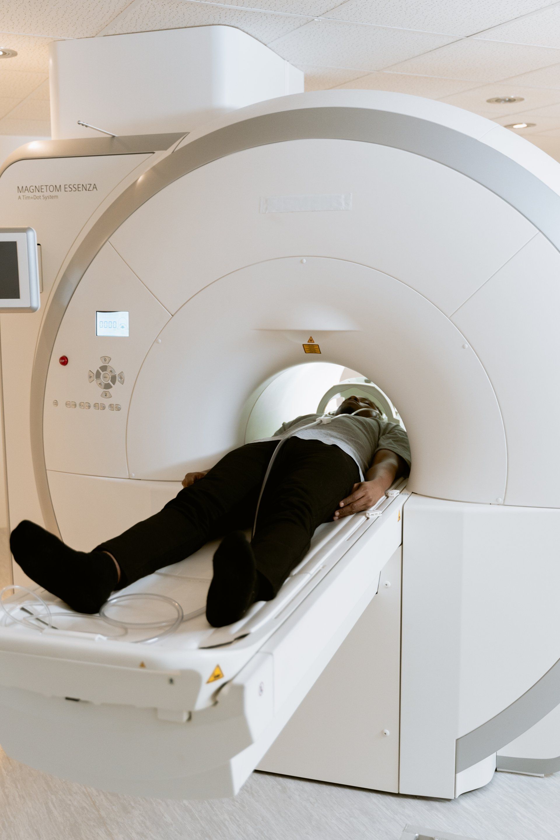
An MRI requires you to lie on a padded table, which then moves into the center of a large enclosed tube-shaped space. If you have issues with claustrophobia, inform your doctor or radiologic technologist so that consideration can be made for mild sedatives.
While the procedure is not painful, you may experience some discomfort if you have some pre-existing back conditions. You will be required to lie still as the images are being taken.
The MRI will produce loud noises, which can be uncomfortable, and you will usually be given earplugs.
To aid with visualizing the images and depending on the reason for the MRI, you may be given an intravenous (IV) contrast dye containing gadolinium. While direct side effects to the dye are minimal, the dye can cause problems if you already have kidney failure or some form of kidney injury. The only scientifically proven adverse effect according to the FDA is a rare condition called nephrogenic systemic fibrosis, which was associated with receiving very high doses of gadolinium in the early 1980-90s.
After the images are taken, the IV will be removed and you will be able to go home right away if the MRI is an outpatient procedure.
Summary
An MRI is a widely used, safe procedure for imaging different tissues and organs that helps your doctor to screen and accurately diagnose conditions such as tumors, infection, or tissue injury.
During the procedure, you will lie still on a table inside a tunnel shaped machine. The scan is not painful, but the MRI machine makes a lot of noise.
Before the MRI, inform your doctor if you have any metal objects or electronic devices in your body such as shrapnel, tooth fillings, pacemakers, or artificial joints. Be sure to remove all metal accessories like jewelry, rings, watches, and glasses.
Sources:
Risk Factors: Radiation - NCI (cancer.gov)
Alzheimer's Disease Fact Sheet | National Institute on Aging (nih.gov)
Nephrogenic Systemic Fibrosis - StatPearls - NCBI Bookshelf (nih.gov)
Gadolinium Side Effects: Toxicity & Nephrogenic Systemic Fibrosis (drugwatch.com)
Gadolinium Contrast Medium (MRI Contrast agents) - InsideRadiology
Multiple Sclerosis | National Institute of Neurological Disorders and Stroke (nih.gov)
Author Bio:
Thandiwe Chavula, MBBS, MSc is a medical doctor and an Immunologist, currently training for a PhD at Baylor College of Medicine in Houston, TX. Her background is in clinical medicine in her home country Malawi. Over the last 6-7 years, her focus and interest has been on translational medical research in autoimmune diseases like Lupus. She works with patient samples and animal models of lupus to further understand disease pathogenesis and discover new treatment targets.
Thank you for reading Patient Education Essentials, the Write Shift RN blog.
Disclaimer: This article was written as a guest post for Write Shift RN LLC's blog. The information in it may not be wholly fact-checked or edited, allowing the reader to see the writer's work and skills firsthand.
This information is not intended as medical advice. It is for informational and educational purposes only. Always talk to your doctor or other qualified healthcare providers about any questions or concerns you may have regarding medical conditions.